|
2025 | Edgar Diaz Miranda, Lei Lei’s lab Confocal micrograph of mouse primary oocytes at the primordial follicle stage. Chemotherapy disrupts the Balbiani body (red), leading to senescence (green-to-blue) and apoptosis (magenta). Instrument: Leica TCS SP8 confocal microscope |
|
2024 | Donielle Brottlund, Felix Fritschi’s lab This image shows a Super Glue epidermal impression of a soybean leaf and is part of a study examining stomatal density. Instrument: Leica Thunder stereomicroscope |
|
2023 | Teka Khan, Michael Roberts’ lab Syncytialization of trophoblasts and localization of the transcription factors SMAD7 (red) and TBX3 (yellow) in syncytial nuclei. Phalloidin (green) was used to mark the cell boundaries. Instrument: Leica TCS SP8 confocal microscope |
|
2022 | Christie Herd, Alexander Franz’s lab La Crosse virus (LACV) can be vertically transmitted from an infected female mosquito to its offspring. This image shows LACV within the developing eggs of Aedes albopictus ovaries, 44 hours after ingestion of a blood-meal containing LACV. Instrument: Leica TCS SP8 confocal microscope |
| 2020 and 2021 | No image contests held. |
|
2019 | Rayne Lim, Shyam Chaurasia’s lab Retinal microvasculature isolated from the mouse eye using trypsin digestion and stained with periodic acid-Schiff stain. Instrument: Zeiss Axiovert 200M widefield microscope |
|
2018 | David Porciani, Donald Burke’s lab When pop art meets a glowing RNA in a dividing B-cell leukemia cell, a fluorescently labeled RNA aptamer is internalized via receptor-mediated endocytosis and transported toward the perinuclear region during endosome maturation. Instrument: Leica TCS SP8 confocal microscope |
|
2017 | Vinit Shanbhag, Michael Petris' lab Focal adhesion and actin cytoskeleton in murine breast cancer cells. Formation of focal contacts and orientation of actin filaments is an important determinant of cell shape, motility and invasiveness. Instrument: Leica TCS SP8 confocal microscope |
|
2016 | Lori Gutzman, Rajiv Mohan’s lab The dermal-epidermal junction in a normal horse hoof. This image is part of a study evaluating differences in collagen between normal horses and those at two different stages of laminitis, since collagen is increased in fibrotic disease. Instrument: Olympus widefield microscope |
|
2015 | Kimberly Jasmer, Mark Hannink's lab Primary normal human embryonic melanocytes immunolabelled for the melanocyte-specific markers Mel5 (Alexa Fluor 488, green) and MART-1 (Alexa Fluor 568, red). Cell nuclei are labeled with DAPI (blue). Instrument: Leica TCS SP8 confocal microscope |
|
2014 | Anju Verma, Melissa Mitchum's lab Cyst nematode feeding sites (syncytia) in Arabidopsis roots. Upon infecting roots, cyst nematodes (dark brown ovals) induce formation of syncytia (swollen roots areas) and feed from them. Normal roots are visible in the background. Instrument: Leica M205 FA stereoscope |
|
2013 | Madeline Miller, Christian Lorson's lab Mouse neuromuscular junctions in a whole-mount neck muscle. The mouse with spinal muscular atrophy (SMN) was treated with morpholinos that correct the pathogenic splicing event in SMN protein, resulting in healthy-looking neuromuscular junctions. Instrument: Zeiss 510 META confocal microscope |
|
2012 | Sunilima Sinha, Michael Roberts' lab Human embryonic stem cells treated for five days with the growth factor BMP4, the inhibitor of activin A signaling A83-01, and the fibroblast growth factor receptor inhibitor PD173074. The cells effectively differentiated to trophoblasts. Instrument: Olympus widefield microscope |
|
2011 | Yuefeng Guan, Shuqun Zhang's lab Selected images from a time-lapse movie showing different stages of an in vitro fertilization in Arabidopsis thaliana. Images are overlays of bright field and YFP fluorescent channels. False colors were added for enhanced visualization. Instrument: Olympus widefield microscope |
|
2010 | Brandie Morgan, Martin Katz's lab Cross section of a motor nerve root taken from the 8th thoracic spinal cord segment in a dog with canine degenerative myelopathy. Each color is a counted nerve fiber. Instrument: Brightfield microscope |
|
2009 | Tom Childs, Frank Booth's lab Co-cultured primary skeletal muscle fibroblasts and muscle precursor cells. Desmin-stained muscle precursor cells (red), α-smooth muscle actin-stained differentiated fibroblasts (green), and DAPI-stained nuclei (blue). Instrument: Olympus widefield microscope |
|
2008 | Yujiang Fang, Helen Mullen's lab Paraffin section of a recipient mouse thyroid 60 days after splenocyte transfer. Transgenic overexpression of the anti-apoptotic molecule FLIP (red) in thyroid epithelial cells (yellow, overlay) promotes resolution of granulomatous experimental autoimmune thyroiditis. Instrument: Zeiss 510 META confocal microscope |
|
2007 | Huachun Wang, Shuqun Zhang's lab Arabidopsis embryo at the heart stage. Green color shows the expression pattern of a GFP marker gene during embryogenesis. The embryo was counterstained with propidium iodide (red) to show the cell outlines. Instrument: Zeiss 510 META confocal microscope |
|
2006 | David Chevalier, John Walker's lab Fluorescent image of GFP expression in the abscission zone of flowers from Arabidopsis thaliana. Instrument: Fluorescent stereomicroscope |
2005 | Katsuhiko Kondo, Bruce McClure's lab Pollen tubes in a self-incompatible Nicotiana alata style stained with aniline blue. Instrument: Fluorescent stereomicroscope |
2004
| Huachun Wang, John Walker's lab Arabidopsis thaliana mature seed showing early torpedo-shaped embryo after clearing with chloral hydrate. The image was obtained using Nomarski optics and processed for 2D deconvolution. Instrument: Zeiss 510 META confocal microscope |

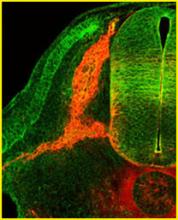
2003
| Jimmy Nail, Mark Kirk's lab |
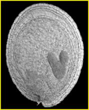
2002
| Bec McClennan, Cathy Krull's lab |
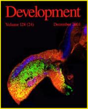
2001
| Mary Swartz, Cathy Krull's lab |