Facility statement
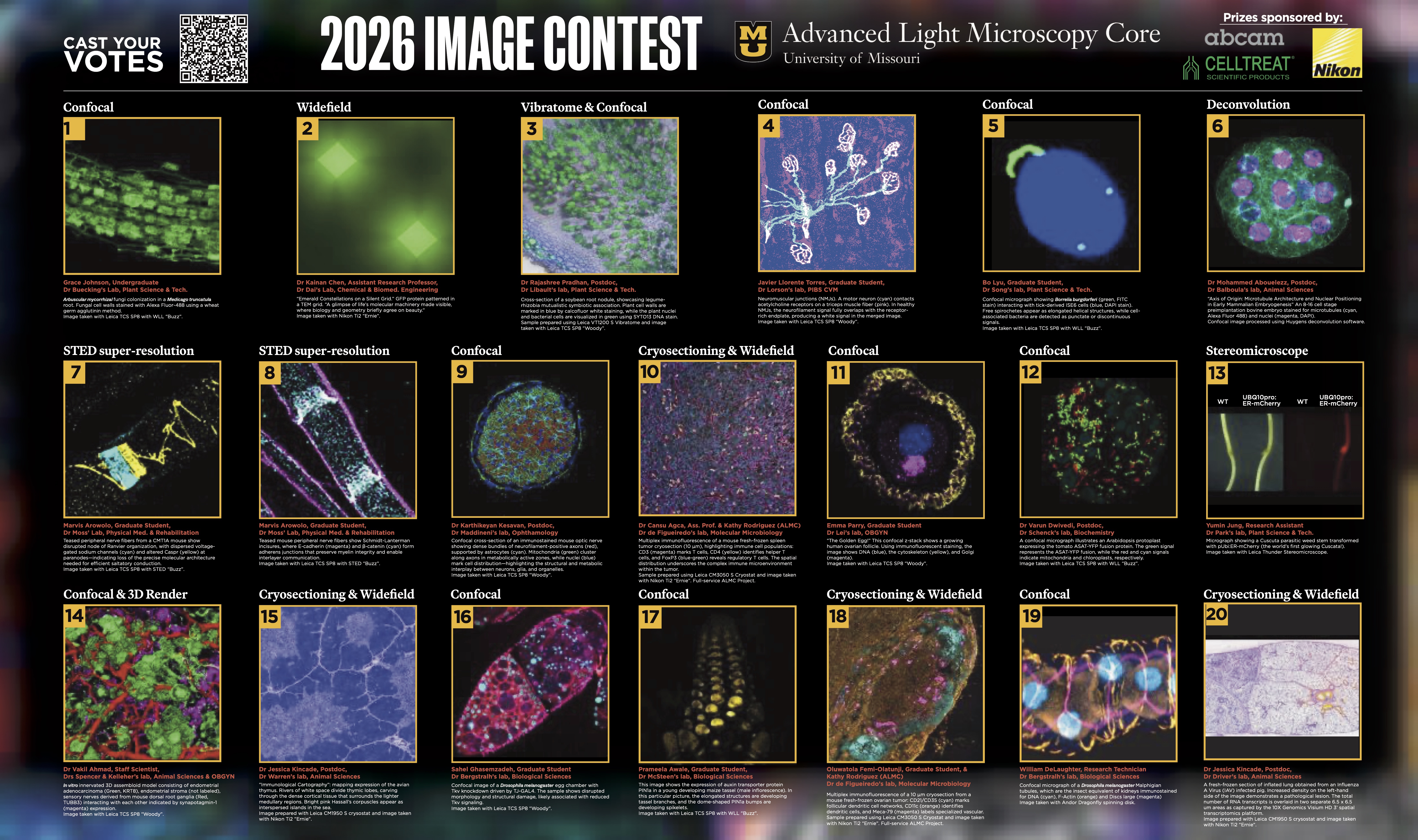
ALMC Image Competition 2026: Voting Now Open!
Winners will be announced during the Show Me Research Week awards ceremony and Closing Reception in Stotler Lounge, Memorial Union North on April 23, 2–3 p.m. for all awards — People's Choice Prize, Expert's Choice Award and the Dr. Alexander Jurkevich Technical Challenge Award.
A poster with all entries can be seen at poster sessions for Show Me Research Week.
Prizes sponsored by Abcam and CellTreat. The People's Choice winner will receive a free Abcam antibody of their choice.
Mission statement
The Advanced Light Microscopy Core (ALMC) is the University of Missouri’s hub for cutting-edge light microscopy, empowering discovery across disciplines. From widefield and confocal to super-resolution and spatial transcriptomics, we provide state-of-the-art imaging technologies, sample preparation equipment, expert guidance, hands-on training and collaborative support. Our mission is to accelerate scientific innovation by making world-class imaging accessible to researchers at Mizzou, partner institutions and industry collaborators throughout the region.
Scheduling
The ALMC is staffed and open 8 a.m.-5 p.m., Monday through Friday year-round excluding university holidays and inclement weather days. Out-of-hours access is available for advanced instrument users.
We use Bookitlab software to manage scheduling and billing. To view calendar availability for our systems and book services, login to Bookitlab with your Mizzou credentials.
To make an appointment on an instrument for the first time, schedule a consultation or arrange training, send your request via email ALMicroscopy@missouri.edu or phone 573-882-4895 during business hours.
About the image: Above is a thin slice of frozen brain tissue — about one-quarter the width of a human hair — from the hippocampus of a cloned pig. The tissue was stained with fluorescent markers to label distinct cell types: astrocytes (green), neurons (red), microglia (purple) and cell nuclei (blue). The confocal image was acquired using an Andor Dragonfly 602 spinning disk confocal microscope, and a 3D reconstruction was generated with Imaris image analysis software. Sample preparation, imaging and analysis were performed using ALMC equipment by Katherine Rodriguez-Lukey, ALMC research specialist. Sample generated as part of the research program of Kiho Lee, associate professor in the Division of Animal Sciences, College of Agriculture, Food and Natural Resources.