Purpose
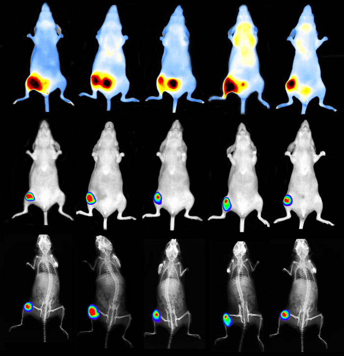
In vivo optical imaging using bioluminescent or fluorescent reporters for the non-invasive study of molecular and biological processes underlying pathophysiology and the development and targeting of novel drug candidates.
Equipment
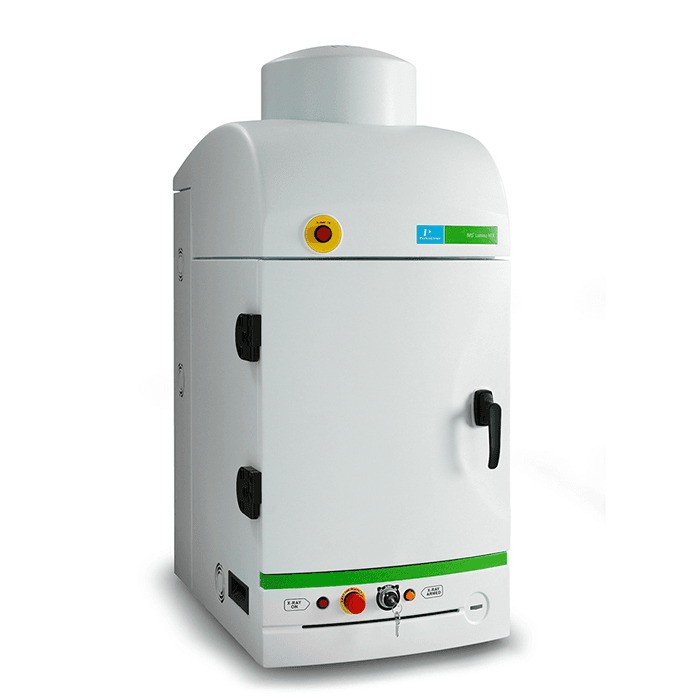
IVIS X5 Lumina Imaging System (Perkin Elmer)
Attached anesthesia unit (RAS-4)
Features
-
High-resolution, low dose X-ray with optical overlay
-
Tunable to image fluorescent sources that emit from green to near-infrared
-
High throughput - 5 mouse field of view
-
Can accommodate small rodents (including rats) to approximately 500g
-
Spectral unmixing for monitoring multiple biological processes
Training and scheduling
Training is required before use. Scheduling is arranged via an online calendar system. You will receive login information after training is completed.
Please review our procedures and contact us for additional information.
Data storage and analysis
Data storage space or devices must be approved before use. All data should be transferred to the user’s approved storage device after an experiment. A designated station can be used for offline data analysis.
Please review our procedures and contact us for additional information.
Costs
Please review our fee schedule.