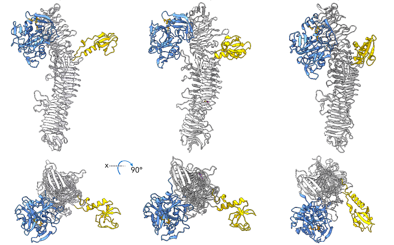
 3D renderings of three bacterial enzymes — EatA, SepA, and Pic — that eat through intestinal mucus to allow infection. Researchers compared their structures to find a shared weakness for potential future vaccine development. Similar structures in the binding part of the enzyme are pictured in blue and the part of mucus cells that bind are pictured in yellow. Images are rendered using AlphaFold 3.
Oct. 30, 2026
By Roger Meissen | Division of Research
Mucus is quite the defense mechanism, and in your gut, it protects from pathogens looking for an opportunity to infect. Yet, everything has a weakness, and bacteria like E. coli and Shigella know how to cut through that mucus lining to create an opening.
But, what if scientists could block the way these infections break through?
For Mizzou’s Zachary Bernsden and David Buckley, that’s their specialty. The structural biologists used Mizzou’s Electron Microscopy Core to examine how human antibodies fight intestinal infections by blocking particular mucus-dissolving proteins made by these bacteria. Their work has helped identify a shared target for future vaccine development that could stop intestinal disease and death for enterotoxigenic E. coli, Shigella and other E. coli strains before infection starts.
“Our bodies mount an adaptive immune response against antigens — molecules our body sees when it’s invaded by a bacteria like E. coli — and these antibodies have a very specific shape that allows them to bind these bacterial antigens to help clear the infection,” said Bernsden, an assistant professor of biochemistry at Mizzou’s School of Medicine. “Our expertise is looking at how antibodies interact with these antigens to uncover the mechanisms they use to block infection.”
Creating a vaccine that protects against multiple intestinal bacteria has long been sought to reduce mortality in less affluent countries. Diarrhea can be deadly under the wrong circumstances even though it’s our immune system’s attempt to flush out invaders and toxins, and in areas with poor sanitation and a lack of dependably clean water, E. coli and Shigella infections lead to hundreds of thousands of deaths a year, especially in children under 5 years old, according to a study in the Lancet.
Bernsden and Buckley teamed up with researchers at Washington University School of Medicine and the International Centre for Diarrheal Disease Research in Bangladesh. The lab of James Fleckenstein, a WashU infectious disease doctor, tested antibodies collected from E. coli-infected Bangladeshi patients and compared them with antibodies from Shigella and other E. coli strains, and they identified several that stop the enzyme secreted by the bacteria — called EatA, SeptA and Pic, respectively — from eating through layers of intestinal mucus.
When samples were handed off, Mizzou’s team took an atomic look.
At Mizzou’s EMC, they plunge their sample into extremely cold liquid ethane to freeze them so fast that ice crystals don’t have time to form. "We take a snapshot of one moment in time," Berndsen explained. Then an electron microscope takes thousands of pictures of the frozen proteins, and a computer combines them into a 3D model.
Through those snapshots, they were surprised to find that the most effective antibodies didn’t block the part of the enzyme that cuts through the mucus, instead blocking the enzyme from productively binding the mucus glycoproteins.
"If you imagine the enzyme like a pair of scissors, you could think, ‘Oh, maybe the antibody will bind on the blade and stop it from cutting,’ but what we found is the antibodies attach far from those blades," Bernsden said. “In a lab test, the antibodies didn't stop the enzyme from snipping a small, simple test molecule but they did stop it from breaking down mucus. The antibodies seem to push the part of the enzyme out of position that grabs the mucus to hold it in place so it can be cut, so the mucus can't line up with the blades.”
Having the accessibility of Mizzou’s EMC helped move this research forward faster.
"You can send samples to other facilities to collect data, but it makes a significant difference being able to collect the data on the fly; We get samples from WashU and within a week can prepare everything we need to collect data on Mizzou’s electron microscope,” said Buckley, a Mizzou biochemistry postdoc. “What really delineates different places are the experts that make up the core, and our EMC is really well staffed, and the training I've gotten there — when I picked up electron microscopy halfway through my PhD — was very substantial.”
Using 3D models showing how the best antibodies attached to the enzymes EatA, SeptA and Pic, they narrowed down the optimal attachment spots that stay nearly identical between all three bacteria.
“We’ve identified three epitopes — three locations on the protein — that are hotspots for different antibodies to bind, but only one of these sites is conserved enough between different bacteria to represent a shared vulnerability,” Buckley said. “So, now we take that information to design a minimal vaccine antigen — the smallest protein that still gets a robust immune response — that directs the immune response to this epitope and nowhere else. The challenge is doing that while still preserving the correct protein shape that allows these antibodies to bind the native protein.”
Now, the Mizzou team has shared their models with their WashU collaborators to make headway toward a potential pan-pathogen vaccine.
“It may not be as lucrative a target as other vaccines, but the military has a lot of interest in it, and bacterial enteric infections cause all sorts of problems,” Bernsden said. “Dr. Fleckenstein is already working with a company to move this work into the clinic, so the ultimate goal is to see if this works in humans.”
This research published June 15, 2026, in Proceedings of the National Academy of Sciences. The work is supported by the National Institute of Allergy and Infectious Diseases (NIAID) of the National Institutes of Health (NIH), and the Department of Veterans Affairs.